
How this tiny animal is helping scientists investigate disorders of the human brain
They’re only an inch long, but the humble zebrafish – a freshwater fish native to south Asia – is allowing scientists to better understand disorders of the human brain and nervous system.
Despite its modest size, the zebrafish’s brain has many similarities to our own, explains Tod Thiele, an assistant professor of biological sciences at U of T Scarborough. The zebrafish “is one of the main genetic animal model systems” he says. “You’ve got the mouse, the fruit fly and roundworms – the zebrafish is right up there with those other animals.”
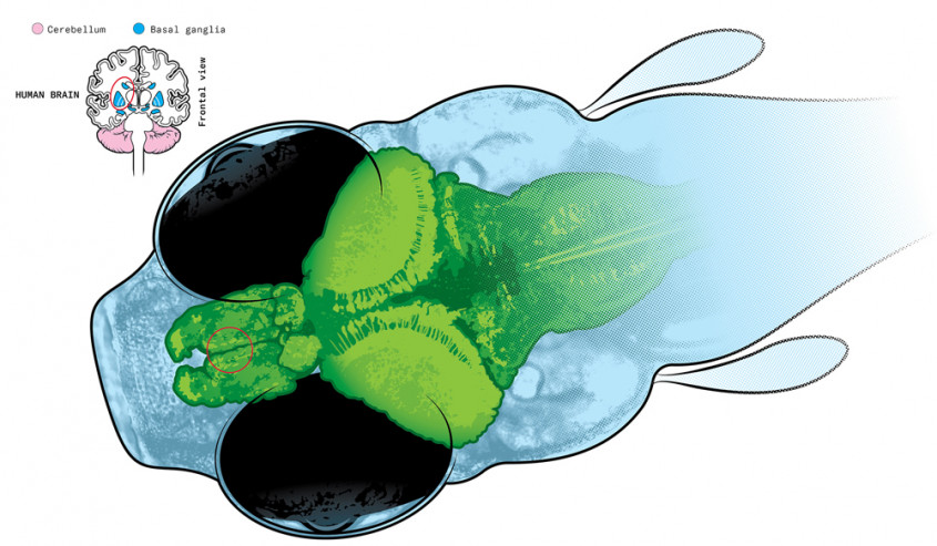
Crucially, the zebrafish is nearly transparent in its early stages, which means researchers can see what all the neurons in the animal’s brain are doing while also monitoring its movements and behaviour. “You don’t have to do a dissection,” says Thiele. “You can just look through the fish.”
Thiele and his colleagues at U of T Scarborough are investigating how the fish’s brain processes sensory information and then uses that information to move about its environment. They are also looking at how those processes are impaired when key neural circuits are damaged. The research could ultimately shed light on movement disorders such as Parkinson’s disease and dystonia, which is characterized by loss of muscle control and involuntary muscle contractions, and can affect people at any age. Parkinson’s is believed to be primarily a disorder of the basal ganglia, while dystonia has been linked to dysfunction in the basal ganglia and the cerebellum – brain regions whose workings can be examined in minute detail in the zebrafish.
The fish is also being studied at Robert Gerlai’s lab at U of T Mississauga and the labs of Ashley Bruce, Henry Krause and Vincent Tropepe at St. George.
WHAT THE LAB IS DOING
Tod Thiele’s lab studies the areas of the zebrafish brain that are evolutionary ancestors to the striatum and pallidum in humans. The lab runs behavioural and imaging experiments on both normal larval zebrafish and ones that have a genetic mutation linked to a hereditary form of dystonia, a movement disorder. By comparing the neuronal activity and movements of the two kinds of fish in minute detail, the scientists hope to gain insight into the brain abnormalities that underlie dystonia.
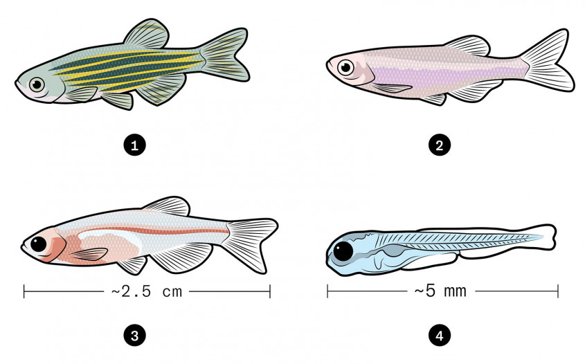
ZEBRAFISH PIGMENT VARIANTS
Albino fish and casper fish have been genetically modified to remove some or all of their skin pigmentation, giving researchers a clearer view of their brain.
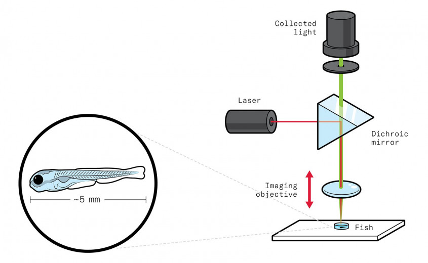
SCANNING THE LARVAL FISH BRAIN
The fish are genetically modified to have a fluorescent protein in all neurons that brightens when a neuron is active. To capture 3D images of brain activity, a low-power infrared laser scans the fish’s brain in a sawtooth pattern while the imaging objective (shown on the left in the illustration below) moves rapidly up and down.
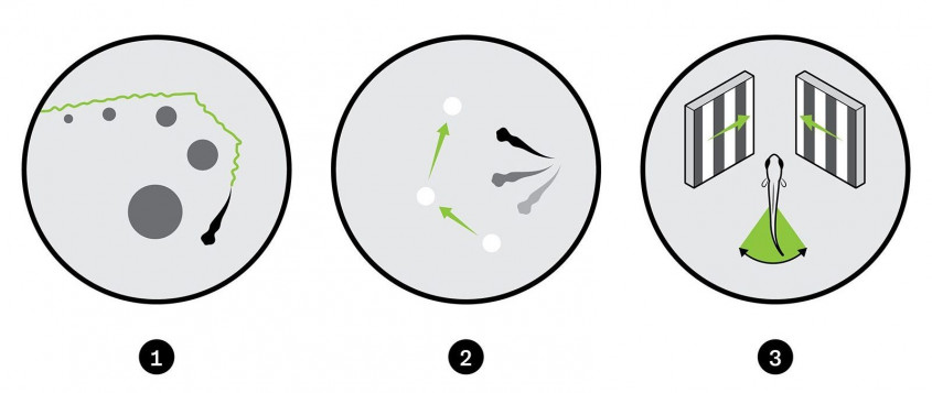
BEHAVIOURAL EXPERIMENTS USING LIGHT
The lab uses three experiments to learn how damage to neural circuits affects the movements of the fish.
This story first appeared in the Winter 2020 issue of U of T Magazine.